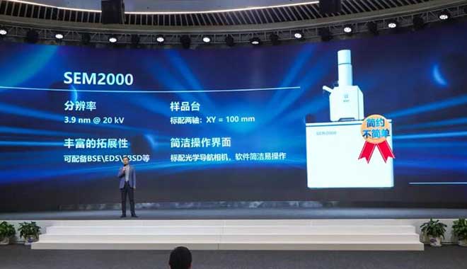
Manufacturing is the mainstay of the real economy, and the importance of manufacturing is being emphasized globally. Scanning electron microscope (SEM) as a powerful analytical instrument, will have a great role in improving manufacturing product innovation and product quality. However, in practice, there are often concerns that the SEM is easily damaged, complicated to use, and takes a long time to get started, resulting in high hidden costs. CIQTEK SEM R&D team to address this pain point, with the goal of "everyone can use" to create an "easy but not simple" tungsten filament SEM2000. CIQTEK tungsten filament scanning electron microscope SEM2000 Easy But Not Simple SEM2000 operation interface is simple, easy to get started, durable, low failure rate and even beginners can easily use it. SEM2000's high degree of automation, key imaging, automatic focus, automatic dispersion, and automatic contrast function, greatly simplify the steps of debugging parameters. SEM2000 has a complete anti-collision process, which can completely avoid the sample from touching the objective lens polar shoe, secondary electronic detector, and other parts. Below are pictures taken by a beginner using SEM2000 after a short training period. Clear image, good contrast, and a large depth of field. If you want to reduce the cost of use and increase the efficiency of operation.If you have never used an electron microscope before and want to try SEM for the first time.If you want the tool to be simpler.Then SEM2000 will be your best choice!
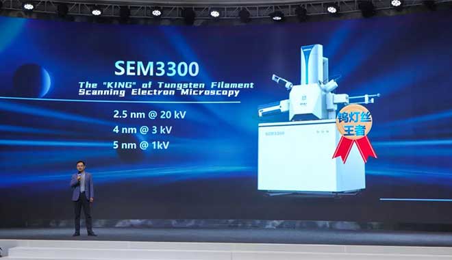
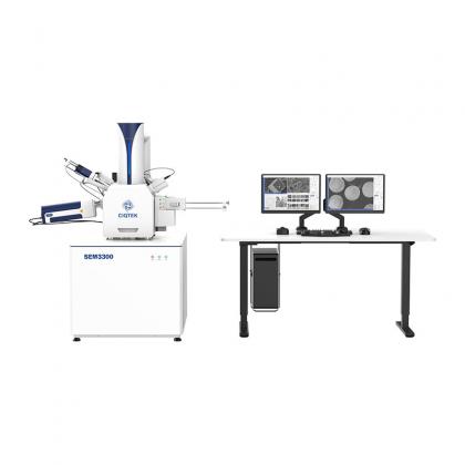
View MoreTungsten filament Scanning Electron Microscopy (SEM) is cost-effective, easy to maintain, relatively simple to operate, and requires less space, making it easy to use by the general public. However, for a long time, the resolution of tungsten filament SEM has been at a standstill, making it difficult to achieve the user's pursuit of higher resolution. CIQTEK recently introduced the SEM3300, a tungsten filament scanning electron microscope that has successfully increased its 20 kV resolution to 2.5 nm, a 16% improvement over ordinary tungsten filament electron microscopes!3 kV resolution of 4 nm, a 2-fold improvement!1 kV resolution of 5 nm, a 3-fold improvement! It redefines the industry standard of tungsten filament scanning electron microscopy by significantly outperforming ordinary tungsten filament electron microscopy in all voltage bands! CIQTEK SEM3300 The following three pictures are real pictures of standard gold particles at different voltages, each particle size is around 300nm, with sharp edges, rich details, and distinct heights. Images of standard gold particles at different voltages taken with SEM3300 It is well known that the diaphragm material in lithium batteries has poor electrical conductivity and tiny pores, and a low-voltage, high-resolution field emission electron microscope must be used to capture better images. Figure a shows the effect of conventional tungsten filament SEM, the details are blurred and unclear. SEM3300 accomplishes this difficult task effortlessly, the septum pores are clearly visible at 1 kV, and the edges of the pores are sharp enough for septum inspection (Figure b). Figure a: Lithium battery septum photographed by conventional tungsten filament SEM, with blurred and unclear details Figure b: SEM3300 photographed lithium battery diaphragm, diaphragm pores clearly visible, the sharp edge of the hole How the CIQTEK SEM3300 is redefining tungsten filament SEM? The CIQTEK SEM R&D team analyzed the main factors limiting the resolution of tungsten filament SEM: The tungsten filament emission structure is a 3-electrode structure with a cathode, gate, and anode. At low acceleration voltage, the filament brightness will be significantly reduced by the space charge effect and electron source aberration. At low landing energy, the chromatic and diffraction aberrations caused by energy dispersion are large, resulting in a large beam spot. In order to ensure the collection efficiency of the lateral secondary electron detector, the working distance is relatively large and the objective magnification is not large enough. In response to these problems, CIQTEK added a 10 kV high voltage tube from the anode directly to the objective lens pole shoe inside the mirror barrel, which we figuratively call a high voltage tunnel. Here is an example of 1 kV landing energy. At the upper end of the high-volta...
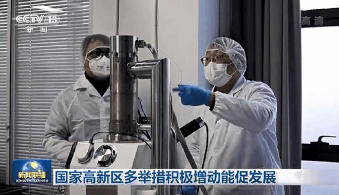
View MoreRecently, China Central Television (CCTV) News interviewed and reported on CIQTEK's scanning electron microscope. "This is a commercial tungsten filament scanning electron microscope with a resolution of 2.5 nm (CIQTEK Tungsten Filament Scanning Electron Microscope SEM3300), which was just released at the end of November." Image source: CCTV NEWS CIQTEK Tungsten Filament Scanning Electron Microscope SEM3300 "In order to be able to move the key core technology from the laboratory to various industries and achieve mass production, a precision measurement technology as the core of the scientific instrumentation industrialization base - Science Instrument Valley (CIQTEK Headquarters Base) is stepping up construction. " Image source: CCTV NEWS Image source: CCTV NEWS
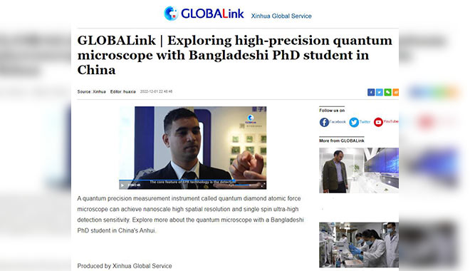
View MoreRecently, CIQTEK was invited by Xinhua News Agency to record a video interview on "Exploring Precision Measurement". CIQTEK is very honored and happy to have this opportunity to spread this technology and information to the public. In the interview, Frank Chen, Overseas Business Development Manager, CIQTEK, demonstrated and introduced the self-developed precision measurement instruments such as the Scanning NV Microscope (SNVM) and Electron Paramagnetic Resonance (EPR or ESR) spectrometer. The following is from Xinhua News Agency (or check the news here): "A precision measurement instrument called Scanning NV Microscope can achieve nanoscale high spatial resolution and single spin ultra-high detection sensitivity. Explore more about it with a Bangladeshi PhD student in China's Anhui. Produced by Xinhua Global Service "
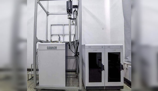
View MoreRecently, the Cryogenic Scanning NV Microscope (SNVM) developed by CIQTEK was successfully delivered to the Institute of Space Environment and Material Science, Harbin Institute of Technology, China. The technical and application engineers of CIQTEK successfully completed the installation and commissioning in the user's laboratory and were highly recognized by the users. CIQTEK staff at the Institute of Space Environment and Material Science of Harbin Institute of Technology adjusted CQDAFM equipment on site Highly sensitive, high spatial resolution precision measurement technology Magnetic properties as one of the fundamental properties of matter, and its microscopic imaging is an important research direction in experimental physics. With the rise of magnetic storage, spintronics, and other fields, the microscopic study of magnetism has put forward completely new technical requirements. In recent years, a unique defect structure Nitrogen-Vacancy (NV) center in diamonds, has attracted the attention of researchers. Precision measurement technology based on the NV center in diamond Scanning probe microscopy based on NV-center in diamond fully combines the advantages of high-sensitivity magnetic detection of light-probing magnetic resonance technology and ultra-high-resolution imaging technology of atomic force microscopy to achieve nanoscale, high-sensitivity, nondestructive, quantitative magnetic properties of scanning imaging, which plays an important role in superconducting magnetic vortex imaging and two-dimensional material magnetic imaging research. Based on the profound technical accumulation in the field of precision measurement, and after extensive research, long-term exploration, and a full understanding of current research needs, the R&D team of CIQTEK has developed the commercialized Cryogenic Scanning NV Microscope. Institute of Space Environment and Material Science, Harbin Institute of Technology, China =The Institute of Space Environment and Material Science of Harbin Institute of Technology is an academic research institute for space physics, space materials, space life, space exploration, and spacecraft application technology. It has built the magnetic substance preparation and comprehensive analysis and testing platform, including a high-throughput ultra-high vacuum magnetron sputtering coating system, multi-chamber pulsed laser molecular beam epitaxial film preparation system, optoelectronic film preparation system, low-temperature Scanning NV Microscope, magnetic force microscope (MFM), magneto-optical Kerr microscope (MOKE), comprehensive physical property testing system (PPMS), etc. Harbin Institute of Technology SNVM | Extraordinary Performance for Frontier Exploration of Magnetic Materials The Cryogenic Scanning NV Microscope is a precision measurement instrument based on the diamond NV center and AFM scanning imaging technolo...
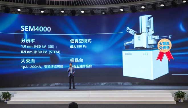
View MoreOn November 24, 2022, CIQTEK successfully held the new electron microscope launch event "Make the Invisible, Visible". Based on the diversified needs of users, CIQTEK launched SEM3300, which "Redefines Tungsten Filament SEM"; SEM2000, which is "Easy But Not Simple"; SEM4000, which is "Super High Beam Current, Super Fast Analysis". >> SEM3300 In the new product launch session, CIQTEK Vice President Feng Cao introduced three new electron microscopes in detail. The first to be unveiled was the SEM3300, a tungsten filament scanning electron microscope, which is a perfect blend of technology and industrial design. With a resolution of 2.5 nm at 20 kV, a 16% improvement over ordinary tungsten filament electron microscopy, a resolution of 4 nm at 3 kV, a 2-fold improvement, and a resolution of 5 nm at 1 kV, a 3-fold improvement, the SEM3300 has significantly surpassed ordinary tungsten filament electron microscopy in all voltage bands, redefining the industry standard for tungsten filament scanning electron microscopy.Take the diaphragm material in lithium batteries as an example, the details of the conventional tungsten filament electron microscopy are blurred and unclear (Figure a below), while the diaphragm pictures taken by SEM3300, the diaphragm pores are clearly visible and the edges of the pores are sharp (Figure b below). Figure a: Li-ion battery septum photographed by conventional tungsten filament electron microscopy, with blurred details. Figure b: SEM3300 photographed lithium battery diaphragm, diaphragm pores clearly visible, sharp edge of the hole. >> SEM2000 Tungsten filament SEM2000 is a product that is not fussy in its operation. It is built with the goal that everyone is able to use it, with a simple interface and rich expandability. If you want a simpler tool, SEM2000 will be your best choice. >> SEM4000 The Field Emission Scanning Electron Microscope SEM4000 is a new product for analytical users' needs. It features a high electron beam current and fast analysis speed, with a maximum electron beam current of over 200 nA and continuously adjustable beam size, which facilitates the selection of the most suitable imaging and energy spectrum conditions. Make achievements for customers, make achievements for peers Dr. Yu He, CEO of CIQTEK, expressed his gratitude to all the guests for their care and support of CIQTEK in his speech. Dr. Yu He said, CIQTEK engraved innovation in its genes, put customers in its heart, insisted on pragmatic innovation with "no compromise", and developed the new product SEM3300 which redefines the industry standard of tungsten filament electron microscope, allowing the tool to return to the tool itself so that users can better rest assured in scientific research. At the end of the conference, CIQTEK Vice President Wan Legend announced the winners of the "Watch Live, Get a Free Electron Microscope (One Year Access)" event. Mr. Zh...
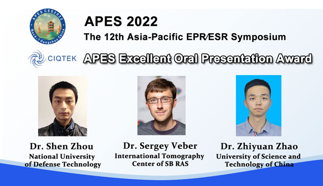
View MoreThe Excellent Oral Presentation Awards are presented during the closing ceremony of the 12th Asia-Pacific EPR Symposium (APES2022) on November 7th, 2022. CIQTEK is pleased to sponsor this award to scientists who have contributed significantly to electron paramagnetic resonance (EPR or ESR) research. This time, congratulations to Dr. Shen Zhou from the National University of Defense Technology, Dr. Sergey Veber from the International Tomography Center of SB RAS, and Dr. Zhiyuan Zhao from the University of Science and Technology of China for winning the awards.APES 2022, Webinar, November 4-7, 2022CIQTEK is happy to sponsor the APES 2022 during November 4-7, 2022. The symposium this year is an online event for international speakers and participants, a new start for Asia-Pacific EPR/ESR Society in the post-epidemic era. The main aims of APES 2022 are to bring together EPR/ESR spectroscopists and to promote and facilitate collaboration among the EPR/ESR community. APES 2022 is intended to stimulate discussions on the forefront of research in all aspects of EPR/ESR ranging from theoretical and experimental advances in CW/Pulsed EPR, high frequency, and high field EPR, ENDOR, PEDLOR/DEER, time-resolved EPR, FMR, MRI, ODMR to applications in medicine, biology, chemistry, materials science and nanotechnology. On November 6, Dr. Sergey Veber gave a presentation entitled "X-band EPR spectrometer based on MW bridge with 300 W solid-state amplifier and AWG unit". Abstract of the PresentationTechnical advances in modern EPR spectrometers set up the frontiers of EPR-related methodologies and approaches. Considering EPR spectrometers of conventional microwave bands, such as X- and Q-, high-power amplifiers, arbitrary wave generators, and fast digitizers are the essential units required for up-to-date pulse EPR techniques.Herein we describe an X-band EPR spectrometer constructed in the Magnetic Resonance Laboratory of Biomolecular Systems (NIOCH SB RAS) and featuring all the required equipment to perform state-of-art pulse EPR experiments. Among the general construction of the spectrometer, the scheme of the microwave bridge is considered in detail including a pulse-forming and pulse-monitoring unit, and a low-noise amplifier with a pulse protection circuit. A modular open-source software "Atomize" (https://github.com/Anatoly1010/Atomize) is used to control the spectrometer including AWG and fast digitizer cards featuring high-speed data streaming. A wideband dielectric EPR resonator was developed to fit the requirements for AWG experiments with chirp pulses. The spectrometer is designed to have a high dynamic range, low coherent noise and to capture the direct dimension efficiently. These capabilities were demonstrated with both rectangular and AWG pulse experiments. This work was supported by the Ministry of Science and Higher Education of the Russian Federation (grant 14.W03.31.0034)Biography of Dr. Sergey VeberDr. Sergey Veber received his Ph.D. in 200...
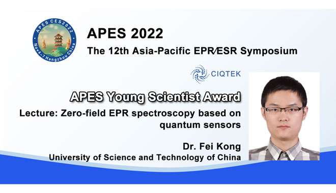
View MoreCIQTEK generously sponsored an annual lectureship and award, given to a young scientist who has made a great contribution to electron paramagnetic resonance (EPR or ESR) research. At the 12th Asia-Pacific EPR/ESR Symposium (APES 2022), the CIQTEK Sponsored Young Scientist Award was awarded to Dr. Fei Kong, CAS Key Laboratory of Microscale Magnetic Resonance and School of Physical Sciences, University of Science and Technology of China.As winner of the 2022 competition, Dr. Fei Kong was invited to gave a prize lecture at the conference, on November 6th.APES 2022, Webinar, November 4-7, 2022CIQTEK is happy to sponsor the APES 2022 during November 4-7, 2022. The symposium this year is an online event for international speakers and participants, a new start for Asia-Pacific EPR/ESR Society in the post-epidemic era. The main aims of APES 2022 are to bring together EPR/ESR spectroscopists and to promote and facilitate collaboration among the EPR/ESR community. APES 2022 is intended to stimulate discussions on the forefront of research in all aspects of EPR/ESR ranging from theoretical and experimental advances in CW/Pulsed EPR, high frequency, and high field EPR, ENDOR, PEDLOR/DEER, time-resolved EPR, FMR, MRI, ODMR to applications in medicine, biology, chemistry, materials science and nanotechnology.Abstract of the Lecture: Zero-field EPR spectroscopyThe nitrogen-vacancy (NV) centers in diamond can promote the sensitivity of EPR detection to a single-spin level even under ambient conditions, and thus enable single-molecule EPR spectroscopy [Science. 347, 1135–1138 (2015); Nat. Methods 15, 697–699 (2018)]. A key figure of single-molecule EPR is that it can resolve heterogeneous molecular information, which is averaged in conventional EPR measurement. However, the current single-molecule measurements are insufficient to do so, due to the poor spectral resolution. Here, I will introduce an easy way to substantially improve the spectral resolution, i.e., removing the magnetic field. The zero-field EPR has been well-studied for decades [Chem. Rev. 83, 49–82 (1983)]. It has narrower spectra with the tradeoff of much poorer sensitivity. Difference from conventional inductive detection, the signal of NV-EPR comes from the statistical fluctuation rather than the thermal polarization of spins, which does not depend on the magnetic field. Therefore, the NV center is an ideal sensor for zero-field EPR detection. I will show that the NV-EPR has the advantage in not only spatial resolution but also spectral resolution.Biography of Dr. Fei KongDr. Fei Kong was born in Hefei, China. In 2012 he obtained a B.Sc. degree at the University of Science and Technology of China (USTC) and later undertook experimental research there on control and simulation using nitrogen-vacancy centers in diamond with Prof. Dr. Jiangfeng Du and Prof. Dr. Fazhan Shi. He received a Ph.D. degree based on this work in 2018. After 3 years of postdoctoral research on t...
View More