Electron-Electron Double Resonance (DEER) in DNA Structure Analysis - EPR (ESR) Applications
Since the 1950s, when Watson and Crick proposed the classical double helix structure of DNA, DNA has been at the heart of life science research. The number of the four bases in DNA and their order of arrangement lead to the diversity of genes, and their spatial structure affects gene expression.
In addition to the traditional DNA double helix structure, studies have identified a special four-stranded DNA structure in human cells, the G-quadruplex, a high-level structure formed by the folding of DNA or RNA rich in tandem repeats of guanine (G), which is particularly high in rapidly dividing G-quadruplexes are particularly abundant in rapidly dividing cells (e.g., cancer cells). Therefore, G-quadruplexes can be used as drug targets in anticancer research. The study of the structure of the G-quadruplex and its binding mode to binding agents is important for the diagnosis and treatment of cancer cells.
Schematic representation of the three-dimensional structure of the G-quadruplex.
Image source: Wikipedia
Electron-Electron Double Resonance (DEER)
The Pulsed Dipolar EPR (PDEPR) method has been developed as a reliable and versatile tool for structure determination in structural and chemical biology, providing distance information at the nanoscale by PDEPR techniques. In G-quadruplex structure studies, the DEER technique combined with site-directed spin labeling (SDSL) can distinguish G-quadruplex dimers of different lengths and reveal the binding pattern of G-quadruplex binding agents to the dimer.
Differentiation of G-quadruplex Dimers of Different Lengths Using DEER Technology
Using Cu(pyridine)4 as a spin label for distance measurement, the tetragonal planar Cu(pyridine)4 complex was covalently bound to the G-quadruplex and the distance between two paramagnetic Cu2+ in the π-stacked G quaternary monomer was measured by detecting dipole-dipole interactions to study the dimer formation.
[Cu2+@A4] (TTLGGG) and [Cu2+@B4] (TLGGGG) are two oligonucleotides with different sequences, where L denotes the ligand. The DEER results of [Cu2+@A4]2 and [Cu2+@B4]2 are shown in Figure 1 and Figure 2. From the DEER results, it can be obtained that in [Cu2+@A4]2 dimers, the average distance of single Cu2+ -Cu2+ is dA=2.55 nm, the G-quadruplex 3′ end forms G-quadruplex dimer by tail-tail stacking, and the gz-axis of two Cu2+ spin labels in G-quadruplex dimer is aligned parallel.
The [Cu2+@A4]2 π stacking distance is longer (dB-dA = 0.66 nm) compared to the [Cu2+@A4]2 dimers. It was confirmed that each [Cu2+@B4] monomer contains an additional G tetramer, a result that is in full agreement with the expected distances. Thus, distance measurements by the DEER technique can distinguish G-quadruplex dimers of different lengths.
Fig. 1 (A) The pulsed EPR differential spectrum (black line) of [Cu2+@A4]2 dimer and its corresponding simulation (red line) (34 GHz, 19 K); (B) After background correction, four phases in a-d DEER time-domain map of the field position (black line) and the best fitting result obtained from PeldorFit (red line); (C) Distance distribution obtained using PeldorFit (red line) and MD simulation (gray line); (D) [Cu2+ Equilibrium between @A4] monomer and [Cu2+@A4]2 dimer. (Angew. Chem. Int. Ed. 2021, 60, 4939-4947)
Fig. 2 (A) DEER time-domain diagrams (black lines) at four field positions a-d after [Cu2+@B4]2 background correction and the best fitting results obtained from PeldorFit (red lines); (B) [Cu2+@B4 ]; (C) Distance distribution obtained using PeldorFit (red line) and MD simulation (gray line). (Angew. Chem. Int. Ed. 2021, 60, 4939-4947)
Probing the binding mode of G-tetramer binding agent to dimer using the DEER technique
Many small molecules and metal complexes, with planar aromatic conjugated systems and positive charges, can bind and stabilize folded secondary structures, thus becoming potential anti-cancer drugs.
N, N ' -bis[2-(1-piperidinyl)ethyl]3,4,9,10-perylenetetracarboxydicarbonyl hydrochloride (PIPER) is a well-known G-quadruplex binding agent that can bind to and stabilize the quadruplex by stacking, and the binding mode of PIPER to the G-quadruplex can be investigated by the DEER technique.
Figure 3 and Figure 4 show the results of DEER experiments with different PIPER to [Cu2+@A4]2 dimer ratios. The results show that when the PIPER to [Cu2+@A4]2 dimer ratio is 1:1 (PIPER@[Cu2+@A4]2), dP = 2.82 nm.
The increased distance between Cu2+-Cu2+ compared to the pure [Cu2+@A4]2 dimers (dA = 2.55 nm) indicates that PIPER forms a sandwich complex with the dimer, with the planar organic molecule interposed between the 3′ faces of the two G tetrameric monomers. When the ratio of PIPER to [Cu2+@A4]2 dimer is 2:1 (2PIPER@[Cu2+@A4]2), d2P = 3.21 nm.
An additional π-stacking distance compared to the PIPER@[Cu2+@A4]2 dimer ( dP = 2.82 nm ) indicates the insertion of two PIPER ligands into the tail-to-tail arranged G-tetramer dimer. The DEER technique can reveal a new binding mode of G -tetramer binding agent PIPER insertion into G-tetramer dimer to form intercalated complexes.
Fig. 3 (A) DEER dipole spectra with different ratios of PIPER and [Cu2+@A4]2 dimer (geff =2.061); (B) DEER modulation with different ratios of PIPER and [Cu2+@A4]2 dimer Depth; (C) Equilibrium of [Cu2+@A4]2 dimer and PIPER@[Cu2+@A4]2, 2PIPER@[Cu2+@A4]2, PIPER@[Cu2+@A4].
(Angew. Chem. Int. Ed. 2021, 60, 4939-4947)
Fig. 4 (A) DEER time-domain spectrum of PIPER@[Cu2+@A4]2; (B) PIPER@[Cu2+@A4]2 distance distribution obtained by using PeldorFit (red line) and MD simulation (gray line); (C) DEER time domain spectrum of 2PIPER@[Cu2+@A4]2; (D) 2PIPER@[Cu2+@A4]2 distance distribution obtained using PeldorFit (red line) and MD simulation (gray line).
(Angew. Chem. Int. Ed. 2021, 60, 4939-4947)
CIQTEK Pulse Electron Paramagnetic Resonance Spectrometer
The CIQTEK pulse electron paramagnetic resonance spectrometer EPR100 supports double electron-electron resonance technology and can be used to study the structural localization, functional interpretation, physiological motion processes, and mechanism of action interpretation of complex membrane proteins, DNA, RNA, nucleic acids-protein complexes, and related protein molecules that have key roles in various diseases.
CIQTEK Pulse Electron Paramagnetic Resonance Spectrometer EPR100
DEER experiment results of CIQTEK EPR100
Experimental results after processing with DeerAnalysis
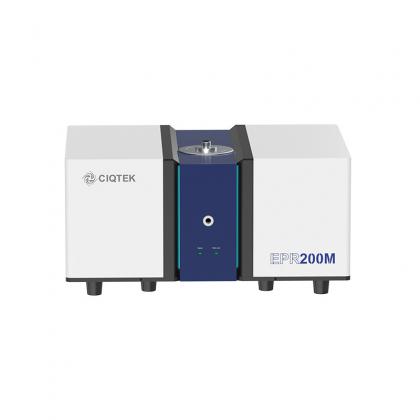
X-band Benchtop Electron Paramagnetic Resonance / Electron Spin Resonance (EPR / ESR) Spectroscopy The CIQTEK EPR200M is a newly designed benchtop EPR spectrometer specializing in the qualitative and quantitative analysis of free radicals, special valence transition metal ions, and material doping and defects. It is an excellent research tool for real-time monitoring of chemical reactions, in-depth evaluation of material properties, and exploration of pollutant degradation mechanisms in environmental science. The EPR200M adopts a compact design and highly integrates the microwave source, magnetic field, probe, and main controller, ensuring sensitivity and stability while being compatible with diverse experimental needs. The user-friendly interface allows even first-time users to start quickly, making this advanced instrument truly easy to use. ★ Email our experts for custom solutions, quotes, or detailed brochures: info@ciqtek.com
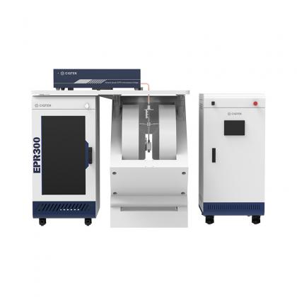
Learn MoreThe CIQTEK EPR200-Plus is a floor-standing EPR spectrometer with enhanced sensitivity for robust continuous wave (CW) EPR measurements. The model can be customized with three different sizes of electromagnets. An upgraded version, CIQTEK EPR300, is another floor-standing CW EPR spectroscopy with higher sensitivity and optional Q-band extension. >> EPR200-Plus Accessories: Dual Mode Resonator, High-temperature System, Liquid Nitrogen Variable Temperature With Cryostat, Liquid helium Variable Temperature, Liquid Helium-free Dry Cryogenic System, Time-resolved EPR System, Goniometers, Irradiation system, Flat cell. Electron paramagnetic resonance (EPR) or electron spin resonance (ESR) spectroscopy is a powerful analytical method to study the structure, dynamics, and spatial distribution of unpaired electronics in paramagnetic substances. It can provide in-situ and non-destructive information on electron spins, orbitals, and nuclei at the microscopic scale. EPR spectroscopy is particularly useful for studying metal complexes or free radicals so it has important applications in the fields of chemistry, materials, physics, environment, etc.
Learn MoreCIQTEK X-band pulse electron paramagnetic resonance (EPR or ESR) spectroscopy EPR100 supports both continuous-wave EPR and pulse EPR functions, satisfying general CW EPR experiments while performing T1 /T2 / ESEEM (electron-spin echo envelope modulation) / HYSCORE (hyperfine sublevel correlation) and other pulsed EPR tests, which can achieve higher spectral resolution and reveal ultra-fine interactions between electrons and nuclei, thus providing users with more information about the structure of matter. >> Optionally equipped with a 4-300 K variable temperature device to enable the detection of paramagnetic substances at ultra-low (high) temperatures.>> EPR100 Accessories: Dual Mode Resonator; High-temperature System; Liquid Nitrogen Variable Temperature With Cryostat; Liquid Helium Variable Temperature; Liquid Helium-free Dry Cryogenic System; Time-resolved EPR System; ELDOR System; ENDOR System; Goniometers; Irradiation System; Flat Cell.
Learn MoreW-band (94 GHz) high-frequency electron paramagnetic resonance (EPR or ESR) spectroscopy compatible with both continuous wave and pulsed EPR test functions EPR-W900 is paired with a slit-type superconducting magnet with a maximum magnetic field of 6 T and can perform variable temperature experiments from 4-300 K.It also has the same software operating platform as the CIQTEK X-band EPR100, providing users with a user-friendly experience.Compared with the traditional X-band EPR technology, high-frequency EPR has many advantages and has important applications in biology, chemistry, and materials.
Learn MoreModernize your old EPR spectroscopy for the cutting-edge EPR research This Modernize will bring you features including: ▶ Higher Sensitivity: Ultra-low noise microwave source and signal detection technology. ▶ Better Resolution: Precise magnetic field control technology ▶ Excellent Compatibility: Compatible with a wide range of EPR spectrometers. ▶ Fast Delivery: Complete delivery of the Modernize hardware within 2-6 months. ▶ High-quality Service: On-site installation and 2-year warranty. ★ Email us for more details: info@ciqtek.com
Learn More