Application of Scanning Electron Microscopy in Failure Analysis of Metallic Materials
Metallic materials are materials with properties such as luster, ductility, easy conductivity, and heat transfer. It is generally divided into two types: ferrous metals and non-ferrous metals. Ferrous metals include iron, chromium, manganese, etc. So far, iron and steel still dominate in the composition of industrial raw materials. Many steel companies and research institutes use the unique advantages of SEM to solve problems encountered in production and to assist in research and development of new products. Scanning electron microscopy with corresponding accessories has become a favorable tool for the steel and metallurgical industry to conduct research and identify problems in the production process. With the increase of SEM resolution and automation, the application of SEM in material analysis and characterization is becoming more and more widespread.
Failure analysis is a new discipline that has been popularized by military enterprises to research scholars and enterprises in recent years. Failure of metal parts can lead to degradation of workpiece performance in minor cases and life safety accidents in major cases. Locating the causes of failure through failure analysis and proposing effective improvement measures are essential steps to ensure safe operation of the project. Therefore, making full use of the advantages of scanning electron microscopy will make a great contribution to the progress of the metal material industry.
01 Electron microscope observation of tensile fracture of metal parts
Fracture always occurs in the weakest part of the metal tissue and records much valuable information about the whole process of fracture, so the observation and study of fracture has always been emphasized in the study of fracture. The morphological analysis of the fracture is used to study some basic problems that lead to the fracture of the material, such as the cause of fracture, the nature of fracture, and the mode of fracture. If we want to study the fracture mechanism of the material in depth, we usually have to analyze the composition of the micro-area on the surface of the fracture, and fracture analysis has now become an important tool for failure analysis of metal components.
Fig. 1 CIQTEK Scanning Electron Microscope SEM3100 tensile fracture morphology
According to the nature of fracture, the fracture can be broadly classified into brittle fracture and plastic fracture. The fracture surface of brittle fracture is usually perpendicular to the tensile stress, and the brittle fracture consists of glossy crystalline bright surface from the macroscopic view; the plastic fracture is usually fibrous with fine dimples on the fracture from the macroscopic view.
The experimental basis of fracture analysis is the direct observation and analysis of the macroscopic morphological and microstructural characteristics of the fracture surface. In many cases, the nature of the fracture, the location of the initiation and the crack extension path can be determined using macroscopic observation, but for a detailed study near the fracture source to analyze the cause of the fracture and the fracture mechanism, microscopic observation is necessary, and because the fracture is an uneven and rough surface, the microscope used to observe the fracture should have the maximum depth of field, the widest possible magnification range and high resolution . Combining these needs, SEM is widely used in the field of fracture analysis. Figure 1 three tensile fracture samples, through low magnification macroscopic observation and high magnification microstructure observation, sample A fracture is a river pattern (Figure A) for typical brittle fracture characteristics; sample B macroscopic no fibrous morphology (Figure B), microstructure no tough nests appear, for brittle fracture; sample C macroscopic fracture consists of glossy facets, so the above tensile fracture are brittle fracture.
02 Electron microscopic observation of steel inclusions
The performance of steel mainly depends on the chemical composition and organization of steel. Inclusions in steel mainly exist in the form of non-metallic compounds, such as oxides, sulfides, nitrides, etc., which cause uneven organization of steel, and their geometry, chemical composition, physical factors, etc., not only make the steel cold and hot processing performance is reduced, but also affect the mechanical properties of the material. The composition, number, shape and distribution of nonmetallic inclusions have a great influence on the strength, plasticity, toughness, fatigue resistance, corrosion resistance and other properties of steel, therefore, nonmetallic inclusions are indispensable items in the metallographic inspection of steel materials. By studying the behavior of inclusions in steel, using the corresponding technology to prevent further formation of inclusions in steel and reduce the inclusions already present in the steel, it is of great importance to produce high purity steel and improve the performance of steel.
Figure 2 Inclusions morphology
Figure 3 TiN-Al2O3 composite inclusions spectral analysis of energy surface
In the case of inclusions shown in Figure 2 and Figure 3, by using SEM to observe the inclusions, together with the energy spectrum analysis of the inclusions contained in the pure iron, it can be seen that the types of inclusions contained in the pure iron are oxide, nitride and composite inclusions.
For example, by measuring the length of the inclusions in the case shown above, it can be seen that the average size of Al2O3 inclusions is about 3 μm, TiN and AlN are within 5 μm, and the size of composite inclusions does not exceed 8 μm; These fine inclusions play a pegging role for the magnetic domains within the electrotechnically pure iron, which will affect the final magnetic properties.
The source of oxide inclusions Al2O3 may be the deoxidation products of steel making and secondary oxides of continuous casting process, the form in the steel material is mostly spherical, a small part of the irregular shape. When observing the inclusions, we should not only observe the morphology and composition of the inclusions, but also pay attention to the size and distribution of the inclusions, which requires a comprehensive evaluation of the inclusions level. For example, if inclusions lead to cracking of the workpiece for failure analysis, large particles of inclusions are usually found at the source of cracking, so it is important to study the size, composition, quantity and shape of inclusions to locate the cause of failure of the workpiece.
03 Scanning electron microscopy method for detecting harmful precipitation phases in steel materials
Precipitated phase is the phase that precipitates when the temperature of saturated solid solution decreases, or the phase that precipitates when the supersaturated solid solution obtained after solid solution treatment is aged, which is a solid-state phase transformation process in which the second phase particles are precipitated and desolvated from the supersaturated solid solution and nucleated. The precipitated phase has a very important role in steel, its strength, toughness, plasticity, fatigue properties and many other important physical and chemical properties have an important impact. Proper control of steel precipitation phase can strengthen the steel properties, if the heat treatment temperature and time control is not appropriate, it will cause a sharp decline in metal properties, such as brittle fracture, easy corrosion, etc.
Fig. 4 Backscatter diagram of SEM3100 pure iron precipitation phase by CIQTEK Scanning Electron Microscope
At a certain accelerating voltage, since the yield of backscattered electrons basically increases with the increase of the atomic number of the specimen, the backscattered electrons can be used as an imaging signal to display the atomic number liner image, and the distribution of chemical components on the surface of the specimen can be observed within a certain range. The atomic number of Pb is 82, and the backscattered electron yield of Pb is high in the backscattered mode, so Pb is bright white in the image.
The hazards of Pb in steel materials are as follows, because Pb and Fe do not generate solid solution, which is difficult to remove in the smelting process, and it is easy to polarize at grain boundaries and form low melting point co-crystals to weaken the grain boundary bonding, so that the hot processing performance of the material is reduced. The possible sources of Pb precipitation in electrotechnically pure iron are the Pb contained in the raw materials of ironmaking and the trace Pb contained in the alloying elements added during smelting. If used for special purposes, the possibility of adding it during smelting is not excluded, with the aim of improving the cutting and processing properties.
04 Conclusion
Scanning electron microscopy as a microscopic analysis tool, can be a variety of forms of observation of metal materials, can be a detailed analysis of various types of defects, metal materials failure of the causes of comprehensive positioning analysis. With the continuous improvement and enhancement of SEM functions, SEM can accomplish more and more work, not only provides a reliable basis for the study of improving material properties, but also plays an important role in the control of production processes, new product design and research.
CIQTEK SEM5000 is a field emission scanning electron microscope(FESEM) with high-resolution imaging and analysis ability, supported by abundant functions, benefits from advanced electron optics column design, with high-pressure electron beam tunnel technology (SuperTunnel), low aberration, and non-immersion objective lens, achieves low voltage high-resolution imaging, the magnetic specimen can also be analyzed. With optical navigation, automated functionalities, carefully designed human-computer interaction user interface, and optimized operation and use process, no matter if you are an expert or not, you can quickly get started and complete high-resolution imaging and analysis work.
Learn MoreStable, Versatile, Flexible, and Efficient The CIQTEK SEM4000X is a stable, versatile, flexible, and efficient field emission scanning electron microscope (FE-SEM). It achieves a resolution of 1.9nm@1.0kV, and easily tackles high-resolution imaging challenges for various types of samples. It can be upgraded with an ultra-beam deceleration mode to enhance low-voltage resolution even further. The microscope utilizes multi-detector technology, with an in-column electron detector (UD) capable of detecting SE and BSE signals while providing high-resolution performance. The chamber-mounted electron detector (LD) incorporates crystal scintillator and photomultiplier tubes, offering higher sensitivity and efficiency, resulting in stereoscopic images with excellent quality. The graphic user interface is user-friendly, featuring automation functions such as automatic brightness & contrast, auto-focus, auto stigmator, and automatic alignment, allowing for rapid capture of ultra-high resolution images.
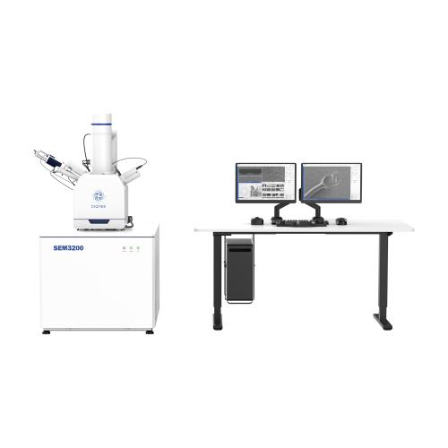
Learn MoreHigh-performance and Universal Tungsten Filament SEM Microscope The CIQTEK SEM3200 SEM Microscope is an excellent general-purpose Tungsten Filament Scanning Electron Microscope (SEM) with outstanding overall capabilities. Its unique Dual-anode electron gun structure ensures high resolution and improves image signal-to-noise ratio at low excitation voltages. Furthermore, it offers a wide range of optional accessories, making the SEM3200 a versatile analytical instrument with excellent expendabilities.
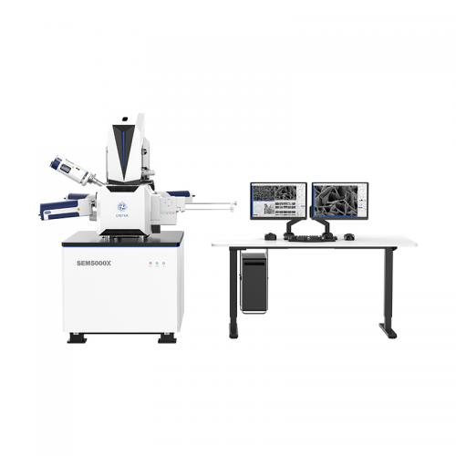
Learn MoreUltra-high Resolution Field Emission Scanning Electron Microscopy (FESEM) Challenges the Limits The CIQTEK SEM5000X is an ultra-high resolution FESEM with optimized electron optics column design, reducing overall aberrations by 30%, achieving ultra-high resolution of 0.6 nm@15 kV and 1.0 nm@1 kV. Its high resolution and stability make it advantageous in advanced nano-structural materials research, as well as the development and manufacturing of high-technology node semiconductor IC chips.
Learn MoreHigh Resolution under Low Excitation The CIQTEK SEM5000Pro is a Schottky field emission scanning electron microscope (FE-SEM) specialized at high resolution even under low excitation voltage. Employing an advanced "Super-Tunnel" electron optics technology facilitates a crossover-free beam path together with an electrostatic-electromagnetic compound lens design. These advancements reduce spatial charging effect, minimize lens aberrations, enhance imaging resolution at low voltage, and achieve a resolution of 1.2 nm at 1 kV, which allows for direct observation of non-conductive or semi-conductive samples, effectively reducing sample irradiation damage.
Learn MoreAnalytical Field Emission Scanning Electron Microscope (FESEM) with Large Beam I CIQTEK SEM4000Pro is an analytical model of FE-SEM, equipped with a high-brightness and long-life Schottky field emission electron gun. The 3-stage electromagnetic lens design offers significant advantages in analytical applications such as EDS / EDX, EBSD, WDS, and more. It comes standard with a low vacuum mode and high-performance low vacuum secondary electron detector, as well as a retractable backscattered electron detector, which benefits the observation of poorly conductive or non-conductive specimens.
Learn MoreHigh-speed Scanning Electron Microscope for Cross-scale Imaging of Large-volume Specimens CIQTEK HEM6000 facilities technologies such as the high-brightness large-beam current electron gun, high-speed electron beam deflection system, high-voltage sample stage deceleration, dynamic optical axis, and immersion electromagnetic & electrostatic combo objective lens to achieve high-speed image acquisition whilst ensuring nano-scale resolution. The automated operation process is designed for applications such as a more efficient and smarter large-area high-resolution imaging workflow. The imaging speed can reach over 5 times faster than a conventional field emission scanning electron microscope (FESEM).
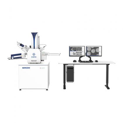
Learn MoreNext-generation Tungsten Filament Scanning Electron Microscope The CIQTEK SEM3300 scanning electron microscope (SEM) incorporates technologies such as "Super-Tunnel" electron optics, inlens electron detectors, and electrostatic & electromagnetic compound objective lens. By applying these technologies to the tungsten filament microscope, the long-standing resolution limit of such SEM is surpassed, enabling the tungsten filament SEM to perform low-voltage analysis tasks previously only achievable with field emission SEMs.
Learn More120kV Field Emission Transmission Electron Microscope (TEM) 1. Divided Workspaces: Users operate TEM in a divided room with comfort reducing environmental interference to TEM. 2. High Operational Efficiency: Designated software integrates highly automated processes, allowing efficient TEM interaction with real-time monitoring. 3. Upgraded Operational Experience: Equipped with a field emission electron gun with a highly automated system. 4. High Expandability: There are sufficient interfaces reserved for users to upgrade to a higher configuration, which meets diverse application requirements.
Learn More