Application of Field Emission Scanning Electron Microscope in Lithium-ion Diaphragm Inspection
I. Lithium-ion battery
The lithium-ion battery is a secondary battery, which mainly relies on lithium ions moving between the positive and negative electrodes to work. During the charging and discharging process, lithium ions are embedded and de-embedded back and forth between the two electrodes through the diaphragm, and the storage and release of lithium-ion energy are achieved through the redox reaction of the electrode material.
Lithium-ion battery mainly consists of positive electrode material, diaphragm, negative electrode material, electrolyte, and other materials. Among them, the diaphragm in the lithium-ion battery plays a role in preventing direct contact between the positive and negative electrodes, and allows the free passage of lithium ions in the electrolyte, providing a microporous channel for lithium ion transport.
The pore size, degree of porosity, uniformity of distribution, and thickness of the lithium-ion battery diaphragm directly affect the diffusion rate and safety of the electrolyte, which has a great impact on the performance of the battery. If the pore size of the diaphragm is too small, the permeability of lithium ions is limited, affecting the transfer performance of lithium ions in the battery, and making the battery resistance increases. If the aperture is too large, the growth of lithium dendrites may pierce the diaphragm, causing accidents such as short circuits or explosions.
Ⅱ. The application of field emission scanning electron microscopy in the detection of lithium diaphragm
The use of scanning electron microscopy can observe the pore size and distribution uniformity of the diaphragm, but also on the multi-layer and coated diaphragm cross-section to measure the thickness of the diaphragm. Conventional commercial diaphragm materials are mostly microporous films prepared from polyolefin materials, including polyethylene (PE), polypropylene (PP) single-layer films, and PP/PE/PP three-layer composite films. Polyolefin polymer materials are insulating and non-conductive, and are very sensitive to electron beams, which can lead to charging effects when observed under high voltage, and the fine structure of polymer diaphragms can be damaged by electron beams. The SEM5000 field emission scanning electron microscope, which is independently developed by GSI, has the capability of low voltage and high resolution, and can directly observe the fine structure of the diaphragm surface at low voltage without damaging the diaphragm.
The diaphragm preparation process is mainly divided into two types of dry and wet methods. The dry method is the melt stretching method, including the unidirectional stretching process and bidirectional stretching process, the process is simple, has low manufacturing costs, and is a common method of lithium-ion battery diaphragm production. The diaphragm prepared by the dry method has flat and long microporous (Figure 1), but the prepared diaphragm is thicker, the microporous uniformity is poor, the pore size and porosity are difficult to control, the assembled battery energy density is low, mainly used in low-end lithium-ion batteries.
Figure 1 Dry stretch diaphragm/0.5KV/Inlens
The wet process, i.e., thermogenic phase separation, involves the mixing and melting of polymers with high-boiling solvents, etc., and the production of microporous membranes through the process of cooling phase separation, stretching, extraction and drying, and heat treatment and shaping. Compared with the dry process, the wet process is stable and controllable, resulting in thin diaphragm thickness, high mechanical strength, uniform pore size distribution and interpenetration (Figure 2). Although the cost of the diaphragm made by the wet process is higher than that of the dry process, the assembled battery has high energy density and good charging and discharging performance, and is mostly used in mid- to high-end lithium-ion batteries. Combined with the pore size analysis system independently developed by GSI, the pore size and porosity of the diaphragm can be analyzed quickly and automatically (Figure 3).
Figure 2 Wet stretch diaphragm/1KV/Inlens
Figure 3 Diaphragm pore size analysis/1KV/Inlens
Although polyolefin-based diaphragms are widely used in lithium-ion batteries, they are limited by the mechanical properties, heat resistance, and surface inertness of the material itself, and simple polyolefin diaphragms cannot meet the requirements of high safety and high performance of lithium-ion batteries. For this reason, surface modification of polyolefin diaphragms is needed to improve their mechanical properties, heat resistance, and affinity with electrolytes. One of the most commonly used methods is the physical coating of the surface of the diaphragm. Inorganic ceramic materials (Figure 4) are characterized by good heat resistance, high chemical stability, and polar functional groups on the surface to improve the wettability of the polyolefin diaphragm to the electrolyte, so they are often used as coated particles to enhance the heat resistance and electrochemical properties of the diaphragm. Figure 5 shows the surface morphology of the ceramic surface of the diaphragm after coating with inorganic ceramic particles.
Figure 4 Alumina ceramic powder/5KV/BSED
Figure 5 Ceramic coated diaphragm/1KV/Inlens
III. Field Emission Scanning Electron Microscope SEM5000
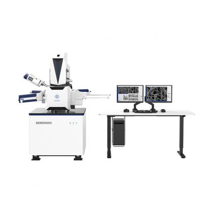
SEM5000 is a high-resolution, feature-rich field emission scanning electron microscope with advanced barrel design, high-voltage tunneling technology, and low aberration non-leakage magnetic objective lens design, to achieve low-voltage high-resolution imaging. Its operating software is equipped with optical navigation to optimize the operation and usage process. Users, whether experienced or not, can quickly get started and complete high-resolution shooting tasks.
CIQTEK SEM5000 is a field emission scanning electron microscope(FESEM) with high-resolution imaging and analysis ability, supported by abundant functions, benefits from advanced electron optics column design, with high-pressure electron beam tunnel technology (SuperTunnel), low aberration, and non-immersion objective lens, achieves low voltage high-resolution imaging, the magnetic specimen can also be analyzed. With optical navigation, automated functionalities, carefully designed human-computer interaction user interface, and optimized operation and use process, no matter if you are an expert or not, you can quickly get started and complete high-resolution imaging and analysis work.
Learn MoreStable, Versatile, Flexible, and Efficient The CIQTEK SEM4000X is a stable, versatile, flexible, and efficient field emission scanning electron microscope (FE-SEM). It achieves a resolution of 1.9nm@1.0kV, and easily tackles high-resolution imaging challenges for various types of samples. It can be upgraded with an ultra-beam deceleration mode to enhance low-voltage resolution even further. The microscope utilizes multi-detector technology, with an in-column electron detector (UD) capable of detecting SE and BSE signals while providing high-resolution performance. The chamber-mounted electron detector (LD) incorporates crystal scintillator and photomultiplier tubes, offering higher sensitivity and efficiency, resulting in stereoscopic images with excellent quality. The graphic user interface is user-friendly, featuring automation functions such as automatic brightness & contrast, auto-focus, auto stigmator, and automatic alignment, allowing for rapid capture of ultra-high resolution images.
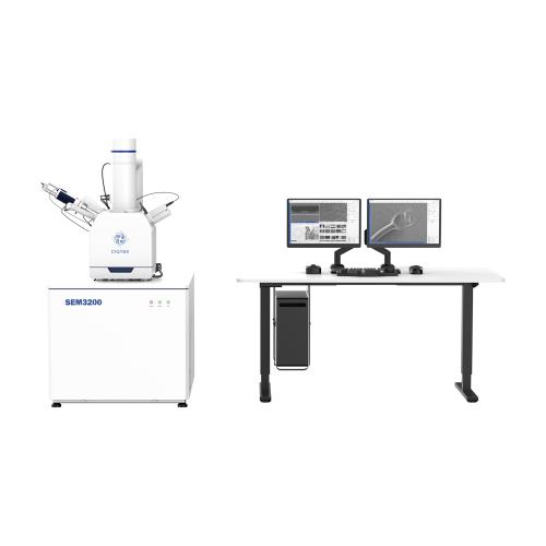
Learn MoreHigh-performance and Universal Tungsten Filament SEM Microscope The CIQTEK SEM3200 SEM Microscope is an excellent general-purpose Tungsten Filament Scanning Electron Microscope (SEM) with outstanding overall capabilities. Its unique Dual-anode electron gun structure ensures high resolution and improves image signal-to-noise ratio at low excitation voltages. Furthermore, it offers a wide range of optional accessories, making the SEM3200 a versatile analytical instrument with excellent expendabilities.
Learn MoreAnalytical Field Emission Scanning Electron Microscope (FESEM) with Large Beam I CIQTEK SEM4000Pro is an analytical model of FE-SEM, equipped with a high-brightness and long-life Schottky field emission electron gun. The 3-stage electromagnetic lens design offers significant advantages in analytical applications such as EDS / EDX, EBSD, WDS, and more. It comes standard with a low vacuum mode and high-performance low vacuum secondary electron detector, as well as a retractable backscattered electron detector, which benefits the observation of poorly conductive or non-conductive specimens.
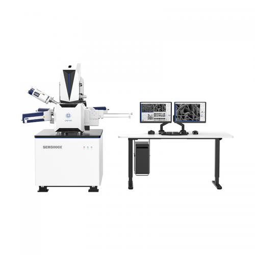
Learn MoreUltra-high Resolution Field Emission Scanning Electron Microscopy (FESEM) Challenges the Limits The CIQTEK SEM5000X is an ultra-high resolution FESEM with optimized electron optics column design, reducing overall aberrations by 30%, achieving ultra-high resolution of 0.6 nm@15 kV and 1.0 nm@1 kV. Its high resolution and stability make it advantageous in advanced nano-structural materials research, as well as the development and manufacturing of high-technology node semiconductor IC chips.
Learn MoreHigh Resolution under Low Excitation The CIQTEK SEM5000Pro is a Schottky field emission scanning electron microscope (FE-SEM) specialized at high resolution even under low excitation voltage. Employing an advanced "Super-Tunnel" electron optics technology facilitates a crossover-free beam path together with an electrostatic-electromagnetic compound lens design. These advancements reduce spatial charging effect, minimize lens aberrations, enhance imaging resolution at low voltage, and achieve a resolution of 1.2 nm at 1 kV, which allows for direct observation of non-conductive or semi-conductive samples, effectively reducing sample irradiation damage.
Learn MoreHigh-speed Scanning Electron Microscope for Cross-scale Imaging of Large-volume Specimens CIQTEK HEM6000 facilities technologies such as the high-brightness large-beam current electron gun, high-speed electron beam deflection system, high-voltage sample stage deceleration, dynamic optical axis, and immersion electromagnetic & electrostatic combo objective lens to achieve high-speed image acquisition whilst ensuring nano-scale resolution. The automated operation process is designed for applications such as a more efficient and smarter large-area high-resolution imaging workflow. The imaging speed can reach over 5 times faster than a conventional field emission scanning electron microscope (FESEM).
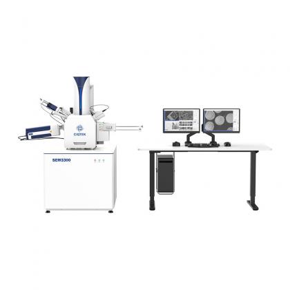
Learn MoreNext-generation Tungsten Filament Scanning Electron Microscope The CIQTEK SEM3300 scanning electron microscope (SEM) incorporates technologies such as "Super-Tunnel" electron optics, inlens electron detectors, and electrostatic & electromagnetic compound objective lens. By applying these technologies into the tungsten filament microscope, the long-standing resolution limit of such SEM is surpassed, enabling the tungsten filament SEM to perform low-voltage analysis tasks previously only achievable with field emission SEMs.
Learn More120kV Field Emission Transmission Electron Microscope (TEM) 1. Divided Workspaces: Users operate TEM in a divided room with comfort reducing environmental interference to TEM. 2. High Operational Efficiency: Designated software integrates highly automated processes, allowing efficient TEM interaction with real-time monitoring. 3. Upgraded Operational Experience: Equipped with a field emission electron gun with a highly automated system. 4. High Expandability: There are sufficient interfaces reserved for users to upgrade to a higher configuration, which meets diverse application requirements.
Learn More